Other Dermatological Conditions
Lumps, bumps, pigment changes, and “not sure what this is?” — doctor-led assessment in Melbourne (Ivanhoe + Diamond Creek)
Not every skin concern fits neatly into acne, rosacea, eczema, or seborrhoeic dermatitis. Many people simply notice a new spot, a changing mole, a persistent bump, or a patch of pigment and want a clear answer.
This page is the hub for “everything else” — common benign lesions and diagnostic uncertainty — with links to relevant condition pages and the best place to start if you’re unsure. (1–3)
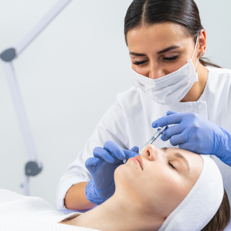
A focused medical assessment with Dr Chris Irwin, including dermatoscopic review where appropriate.
Key takeaways
- Many skin lesions look similar — accurate diagnosis matters before treatment or removal. (1–3)
- Some lesions are harmless but annoying; others need monitoring or biopsy for certainty. (1–3)
- If you’re not sure what you have, a 20 minute doctor appointment is the fastest pathway.
Jump links
- When to book promptly
- What we can help with
- Common “other” conditions (linked pages)
- What happens at your appointment
- Removal options (when appropriate)
- FAQs
- Book
When to book promptly
Book promptly if you notice:
- A new or changing mole (size, colour, shape, bleeding, itching). (3)
- A spot that is growing quickly, crusting, or bleeding
- A lesion that looks “stuck on” but is changing or irritated
- Any lesion you feel uncertain about — especially if you have higher skin cancer risk factors. (3)
What we can help with
This hub covers:
- Moles and atypical/dysplastic naevi (monitoring vs removal for certainty). (2)
- Benign “stuck-on” growths and pigment spots. (1–3)
- Viral lesions (including warts and molluscum). (5)
- Fibrous bumps (e.g., dermatofibroma)
- Small cysts and superficial lumps
- General diagnostic uncertainty — when you just want a clear answer
Common “other” conditions (linked pages)
Pigment and mole-related
- Dysplastic naevi (HL48)
“Atypical moles” that may need monitoring or removal depending on features and risk profile. (2) - Harmless moles / naevi (HL56)
Reassurance, monitoring strategies, and when removal is sensible for irritation or cosmetic reasons. - Solar lentigo (HL50)
Sun-related pigment spots (“sun spots”) and how we assess and treat them. (1) - Skin cancer types and information (HL80)
If your concern is a changing spot, bleeding lesion, or non-healing area, start here. (3)
Lumps and bumps
- Seborrhoeic keratosis (HL51)
Very common benign “stuck on” lesions that can mimic more serious conditions when inflamed or irritated. (1,4) - Dermatofibroma (HL55)
Firm benign nodules (often on the limbs) that can be tethered and persistent. - Milia (HL57)
Small superficial cysts (often around the eyes/cheeks) and safe treatment options. - Lichenoid Planus Like Keratosis (LPLK) (HL52)
A regressing inflammatory lesion that can mimic other pigment changes clinically and on dermoscopy.
Viral and contagious lesions
- Warts (HL53)
Common viral lesions with multiple treatment options depending on location, size, and patient preference. - Molluscum contagiosum (HL54)
Common in children; management depends on extent, symptoms, and eczema overlap. (5)
Texture and “bumpy skin”
- Keratosis pilaris (HL49)
“Chicken skin” bumps caused by keratin plugging follicles — harmless but often cosmetically bothersome. (6)
What happens at your appointment (20 minutes)
- A focused medical assessment with Dr Chris Irwin
- Dermatoscopic review where appropriate
- A clear diagnosis and an “is this concerning?” answer
- A plan: monitor, treat, remove, biopsy, or refer — depending on the lesion
Removal options (when appropriate)
For benign lesions, removal is sometimes chosen because:
- the lesion is irritating (snagging, bleeding, inflamed)
- histology is needed for certainty
- removal offers the best cosmetic pathway (depending on lesion type, depth and location)
Options may include shave + base treatment, laser, radiofrequency, cryotherapy, or surgical excision — chosen on medical appropriateness and cosmetic outcome.
FAQs
Frequently Asked Questions
If it’s benign, should I still remove it?
Not necessarily. Many benign lesions can be monitored. Removal is often about symptoms (irritation), cosmetics, or diagnostic certainty. (1–3)
If I’m not sure what it is, which page should I start with?
Start here and book a 20 minute appointment. If your concern is a changing spot, also see Skin cancer types and information. (3)
Do you send lesions for histology?
Almost always, yes. We believe this is the safest and most thorough approach.
The main exception is when a lesion is destroyed in a way that leaves no tissue to send (for example, some laser treatments or cryotherapy). Even in these situations, to be as thorough as possible, Dr Irwin will sometimes take a gentle shave sample first and send that for histology before completing the ablation.
Let's start
Book
If you have a lump, bump, mole, or pigment change and you’d like a clear diagnosis and a sensible plan:
Ivanhoe: Unit 1, 1065 Heidelberg Road, Ivanhoe VIC 3079
Diamond Creek: Shop 12, 67 Main Hurstbridge Road, Diamond Creek VIC 3089
References
1.DermNet NZ. Common benign skin lesions. https://dermnetnz.org/topics/benign-skin-lesions
2.DermNet NZ. Atypical naevi (dysplastic nevi). https://dermnetnz.org/topics/atypical-melanocytic-naevus
3.Australasian College of Dermatologists. Skin cancer information. https://www.dermcoll.edu.au/skin-cancer/
4.Australasian College of Dermatologists. Seborrhoeic keratoses. https://www.dermcoll.edu.au/atoz/seborrhoeic-keratoses/
5.Australasian College of Dermatologists. Molluscum contagiosum. https://www.dermcoll.edu.au/atoz/molluscum-contagiosum/
6.DermNet NZ. Keratosis pilaris. https://dermnetnz.org/topics/keratosis-pilaris